Fig 1.

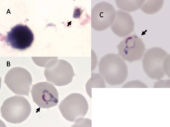
Giemsa staining of P. knowlesi parasites in thick (A) and thin (B and C) films prepared on the day of hospital admission. Arrows point to parasites. Films and stains were prepared by standard techniques. Magnification, ×1,000.

Giemsa staining of P. knowlesi parasites in thick (A) and thin (B and C) films prepared on the day of hospital admission. Arrows point to parasites. Films and stains were prepared by standard techniques. Magnification, ×1,000.