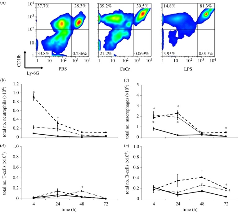
Figure 4.
A flow cytometric analysis of the inflammatory exudate recovered at different time points. (a) Identification of cells within the inflammatory exudate recovered 4 h post implantation; cells that were Ly-6G+ and CD11b+ were determined to be neutrophils, Ly-6G− and CD11b+ cells were identified as monocytes/macrophages. (b) Number of neutrophils, (c) monocytes/macrophages, (d) T-lymphocytes and (e) B-lymphocytes present in the inflammatory exudates at different time points following injection of PBS (solid line with squares), CoCr (dotted line with diamonds) debris or LPS (dashed line with circles). Results are means ± s.e.m. (n = 3). Asterisks (*) denote significantly different from PBS (negative control) values (p < 0.05) by one-way ANOVA followed by Dunnett's multiple comparison test. (Online version in colour.)

