Figure 1.
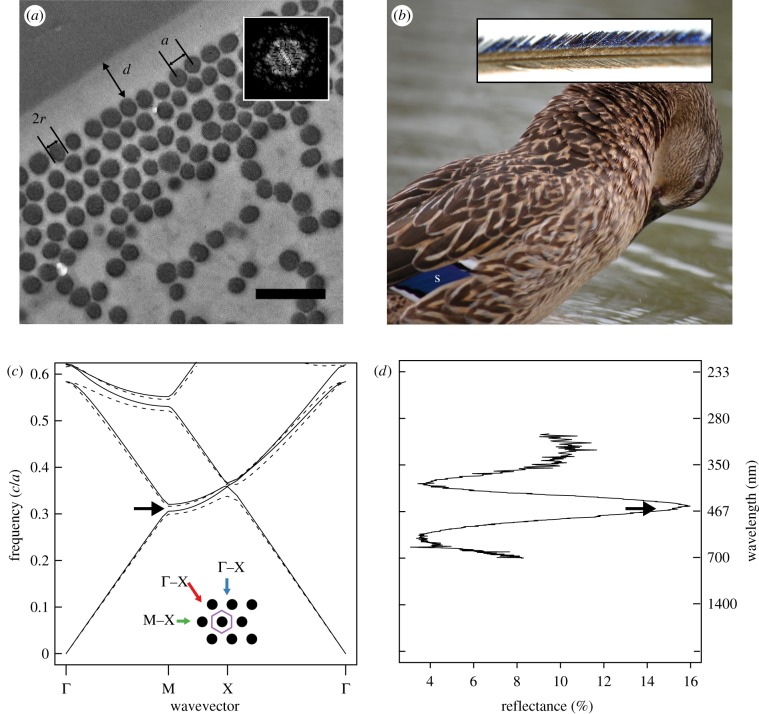
Feather nanostructure and colour-producing mechanism in iridescent duck feathers. (a) Representative cross section of an iridescent feather barbule taken from the speculum feather of a mallard (A. platyrhynchos) showing measured parameters a (lattice constant), 2r (melanosome diameter) and d (cortex thickness). Inset shows Fourier transformation of region pictured in (a), illustrating hexagonal periodicity of melanosomes. (b) Photograph of a female mallard showing location of speculum (s) and light microscope image of a feather barb (inset). (c) Band structure diagram of two-dimensional hexagonal lattice of melanin rods (r/a = 0.44; RI = 2.0) embedded in keratin (RI = 1.56); lines represent modes for s- (E-field parallel to rods; dashed lines) and p-polarized light (E-field perpendicular to rods; solid lines), and black arrow shows location of partial photonic band gap (PBG). Inset shows schematic diagram of barbule cross section (unit cell, purple hexagon), depicting light incident along Γ–M (blue arrow), M–X (green arrow) and X–Γ directions (red arrow). (d) Plot of feather reflectance at corresponding frequencies in (c) with wavelength shown on secondary y-axis; black arrow shows location of partial PBG as in (c).

