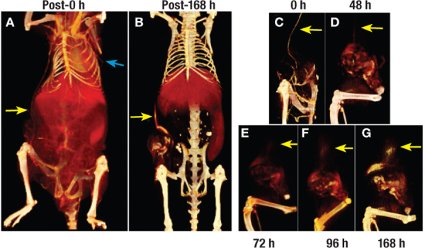
Figure 7.
Functional imaging of tumor vasculature. Visualization of co-opted (yellow arrow) and intratumoral vasculature in a mouse model of breast cancer. Longitudinal imaging enabled functional evaluation of both intratumoral and extratumoral blood vessels. The highly permeable nature of co-opted tumor blood vessel located outside the tumor margins resulted in diffuse signal enhancement due to extravasation of liposomal contrast agent into the perivascular space (yellow arrow).32