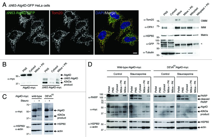
Figure 2. Import of ΔN63 Atg4D into the mitochondrial matrix does not depend upon caspase action. (A) Mitochondrial targeting of ΔN63 Atg4D-GFP. To the left, example field of HeLa cells stably expressing ΔN63 Atg4D-GFP and stained with Tom20 antibodies (Bar = 10 µm). To the right, immunoblots of ΔN63 Atg4D-GFP stable HeLa cell extracts fractionated into post nuclear supernatant (PNS), cytosol, mitochondria (mitos) and mitoplasts (PK: proteinase K). Arrow on the anti-GFP blot indicates the position of ΔN63 Atg4D-GFP. (OMM: outer mitochondrial membrane; IMM: inner mitochondrial membrane). (B) Immunoblot of PNS, cytosol, and PK-treated mitochondrial fractions of HeLa cells transiently expressing Atg4D-myc or ΔN63 Atg4D-myc. The ~42 kDa mitochondrial form of Atg4D is indicated (* denotes an intermediate band resolving between ΔN63 Atg4D-myc and the ~42 kDa mitochondrial product). (C) Immunoblots of lysates of HeLa cells transiently expressing wild-type or caspase-resistant (DEVA63) Atg4D-myc, incubated in the absence or presence of staurosporine (6 h). (C) Immunoblots of fractionated HeLa cells expressing Atg4D-myc or ΔN63 Atg4D-myc, incubated in the absence or presence of staurosporine (6 h).

An official website of the United States government
Here's how you know
Official websites use .gov
A
.gov website belongs to an official
government organization in the United States.
Secure .gov websites use HTTPS
A lock (
) or https:// means you've safely
connected to the .gov website. Share sensitive
information only on official, secure websites.
