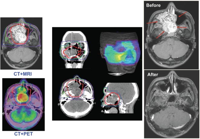
Figure 2.
In treatment planning, fusion images using the computed tomography, magnetic resonance imaging and positron emission tomography are commonly used (left). Dose distributions of the different planes and facial surface (middle). The patient with adenoid cystic cancer before and after carbon ion radiotherapy (right).

