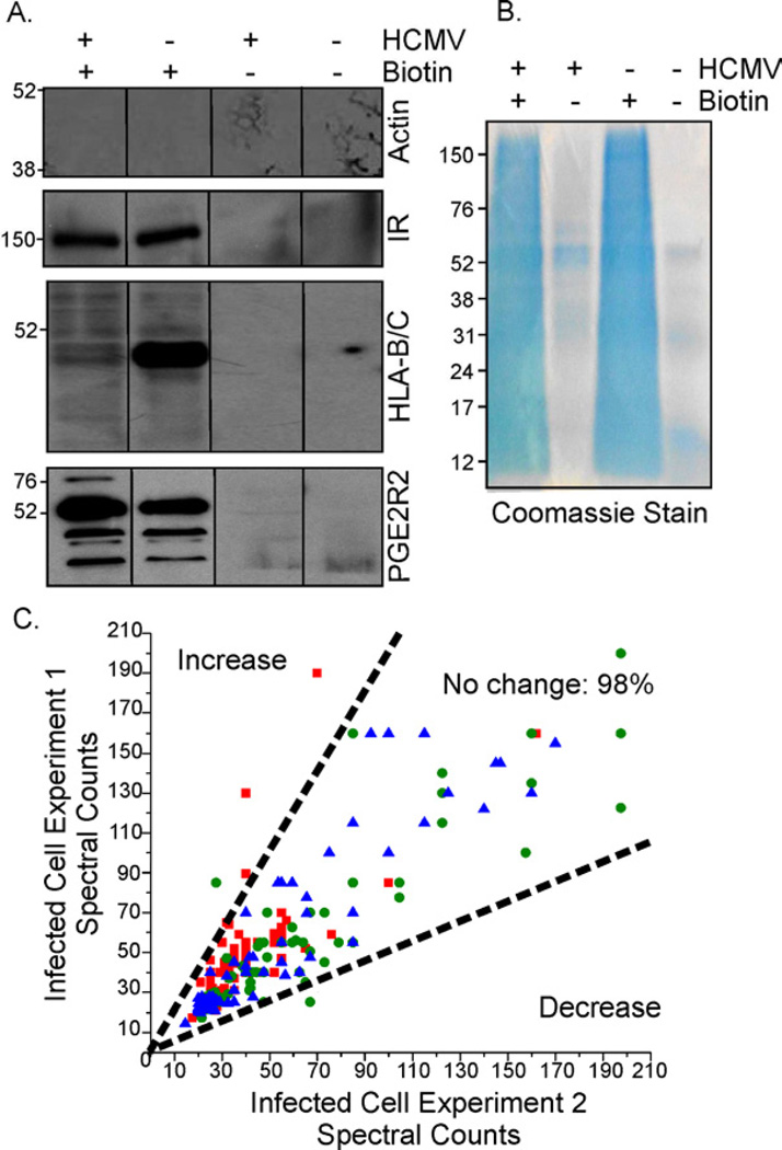
Fig. 1. Identification of cell surface protein alterations in HCMV-infected cells.
Fibroblasts were mock-infected or infected at a multiplicity of 5 IU/cell.
(A) Specificity of biotin labeling: Avidin-purified cell surface protein preparations were assayed by Western blot for β-actin, insulin receptor (IR), MHC-class I receptor (HLA-B/C), and prostaglandin receptor 2 (PGE2R2).
(B) Purification of biotinylated proteins: Avidin-purified samples (~50 µg protein in +biotin samples) were subjected to electrophoresis and Coomassie stained.
(C) Reproducibility of MS data across independent experiments: Normalized spectral counts of infected cell proteins isolated at 6 (■), 24 (●) and 72 hpi (▲) were plotted from two independent experiments. Dotted-lines mark a 2-fold difference in counts.

