
Figure 1. Growth, proliferation, and recapitulation of stem cell niche-like microenvironment of cells under cardiosphere culture condition.
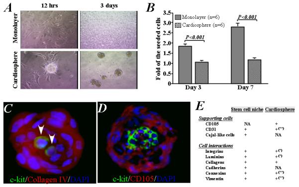
A) Representative images show the growth of cardiac stem cells into cell aggregates and suspended cardiospheres on poly-D-lysine-coated plate (left) or as adherent monolayer cells on a fibronectin-coated plate (right). B) The number of cells increased 2- and 3-fold after 3 and 7 days in monolayer culture, while proliferation was lower in cardiosphere culture. C) Representative images show that two c-kit-positive (green) cardiac stem cells in the central area of a cardiosphere with abundance of collagen IV (red). D) A cluster of c-kit-positive stem cells is localized in the central core of cardiospheres, surrounded by CD105-positive supporting cells. E) Tabular summary of features of cardiac stem cell niches and cardiospheres [NA: not assessed; *based on data presented in Supporting Information Figure 6; ** based on our previous data (Ref #25)].