
Figure 5. Engraftment of human cardiac-derived cells 3 weeks after implantation into the infarcted hearts of mice.
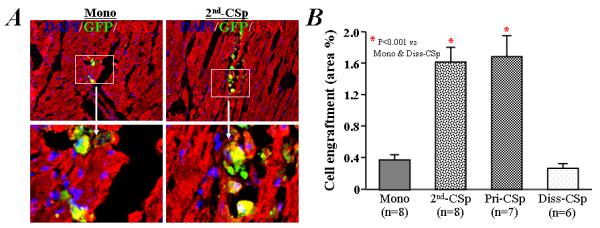
A) Engraftment of GFP+ human cells was infrequently observed in mice receiving the implantation of single cell suspension of CDCs after 3 days culture under monolayer condition (left). However, the survival of GFP+ human cardiac-derived cells was more evident in mice receiving a portion of the same initial CDCs after 3 days of culture under cardiosphere condition (2nd-CSps, right). α-SA: α-sarcomeric actin. B) Quantitative data for cell engraftment (percentage of green (GFP+) area/total area) in the infarcted heart after implantation.