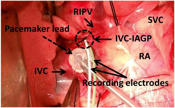
Figure 1.
One pair of electrodes under fascia of inferior vena cava – inferior atrial ganglionated plexus (IVC-IAGP, circle) and an epicardial pacemaker lead (dashed arrow) at the junction of right atrium (RA), left atrium and IVC. IVC-IAGP is located at the junction of the inferior vena cava and left atrium. Because IVC-IAGP electrodes are located at the atrioventricular junction, a local ventricular electrogram is always recorded to allow VR analyses. The relative sizes of A, V and T waves vary depending on the location of the electrodes and the filter settings. RIPV; right inferior pulmonary vein, SVC; superior vena cava, IVC; inferior vena cava.

