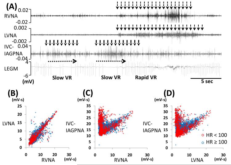
Figure 5.
Slowing of VR associated with IVC-IAGPNA discharge without RVNA or LVNA discharge. Figure A shows independent IVC-IAGPNA resulted in reduction of VR while RVNA and LVNA discharge without IVC-IAGPNA discharge was associated with rapid VR. One dog showed a linear relation between LVNA and RVNA (B) and an ‘L’-shaped relation between RVNA, LVNA with IVC-IAGPNA (C, D). B shows that RVNA and LVNA showed linear relation when VR≥ 100 bpm (blue dots). Unlike that shown in Figure 4, the panels C and D show that blue and red dots are observed at both arms of the L-shaped correlation.

