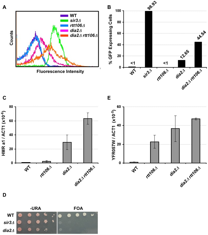
Figure 1. Dia2 functions in parallel with Rtt106 in HMR silencing.
(A–C) Mutant dia2Δ cells have defects in transcriptional silencing at the HMR locus. Cells with the indicated genotype were analyzed for GFP expression using flow cytometry (A), and the percentage of cells expressing GFP was reported (B). Wild-type (WT) cells (less than 1%) and cells with SIR3 deleted (sir3Δ, over 95%) were used as standards to set the gate for GFP expression. The flow cytometry profile and percentage of cells expressing GFP are reported from one of three independent experiments. (C) HMR a1 gene expression is elevated in dia2Δ and dia2Δ rtt106Δ mutants. RNA was collected from cells of the indicated genotype and reverse transcribed. Expression of the HMR a1 gene was analyzed via real-time PCR, normalized against the expression of the ACT1 gene and reported as the average ± standard deviation (s.d.) of two independent experiments. (D–E) Telomere silencing is compromised in dia2Δ cells. (D) Telomere silencing was assayed using the URA3 gene integrated at the left end telomere of chromosome VII and growth on medium containing 5-fluoroorotic acid (FOA). (E) YFR057W expression levels were determined by real time RT-PCR and analyzed as described above with the average ± s.d. of two independent experiments shown.

