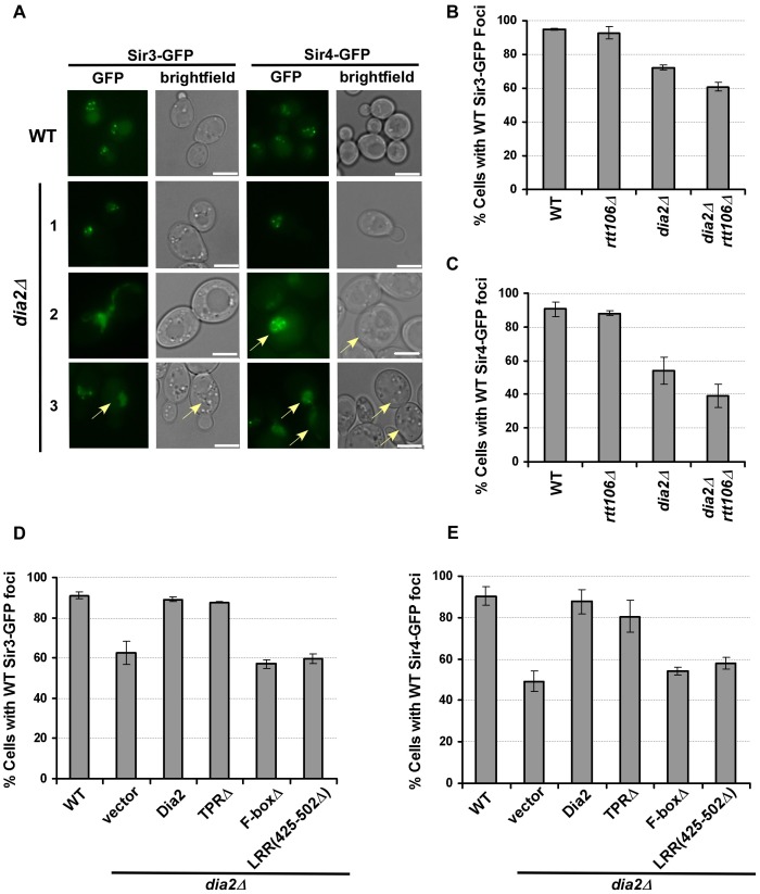
Figure 4. Sir3 and Sir4 are mislocalized in dia2Δ cells and cells expressing dia2 mutants lacking the Dia2 F-box or LRR domain.
(A–C) Sir3 and Sir4 are mislocalized in dia2Δ and dia2Δ rtt106Δ cells. (A) Representative images of Sir3-GFP or Sir4-GFP foci in WT and dia2Δ cells. Cells expressing Sir3-GFP and GFP-Sir4 exhibited nuclear foci in WT cells, and this pattern is lost in some dia2Δ cells. Note that dia2Δ cells are larger in size than wild-type cells (images were taken under the same magnification). The scale bar represents 5 µm. Cells that exhibited Sir protein localization defects as described in the text were marked by arrows. (B–C) Quantification of the percentage of dia2Δ cells that had WT-like Sir3-GFP foci (B) and WT-like Sir4-GFP foci (C). (D–E) Cells expressing dia2 mutants lacking the Dia2 F-box and LRR domains exhibit defects in the localization of Sir3 (D) and Sir4 (E). For each experiment, at least 100 cells were counted for each genotype, and the average ± s.d. percentage of cells expressing WT-like foci from at least two independent experiments was reported.

