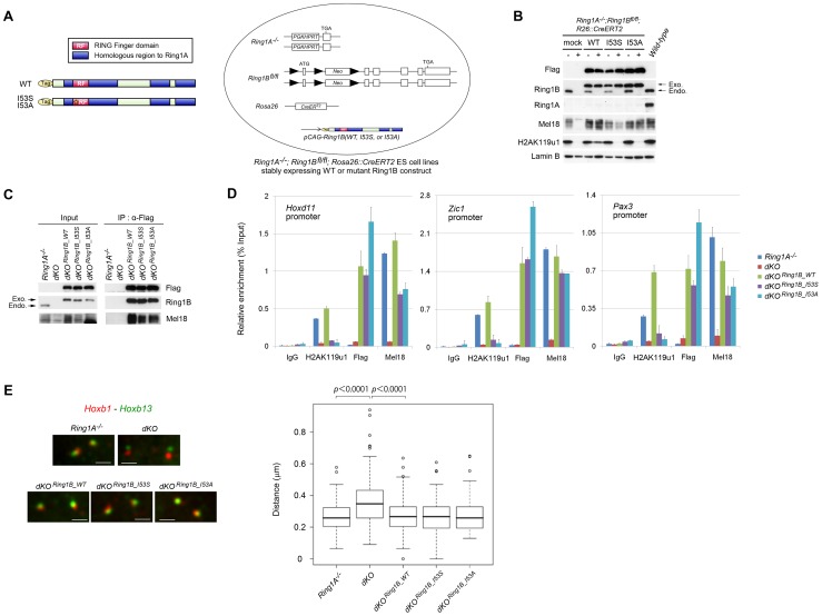
Figure 2. Generation of ESCs expressing catalytically inactive Ring1B.
(A) Schematic representation of 3xFlag-tagged Ring1B, showing wild-type and point-mutant derivatives. Each of these construct was stably transfected into Ring1A−/−; Ring1Bfl/fl; R26::CreERT2 ESCs. (B) Immunoblot analysis of Ring1A, Ring1B, Flag, H2AK119u1 and Lamin B protein levels in whole cell lysates of wild-type and Ring1A−/−; Ring1Bfl/fl; R26::CreERT2 ESC lines expressing mock, WT, I53S, or I53A Ring1B with or without OHT treatment (OHT+ and −, respectively). (C) Immunoprecipitation (IP) analysis showing the association of exogenous Ring1B WT, I53S or I53A with an endogenous PRC1 component Mel18. Extracts of OHT-untreated (−) and -treated [(+); day 2] Ring1A−/−; Ring1Bfl/fl; R26::CreERT2 ESC lines expressing each of the constructs were immunoprecipitated with anti-Flag antibody. Resulting precipitates (IP) and lysates (Input) were immunoblotted with antibodies against Flag, Ring1B and Mel18. (D) Association of Flag-tagged proteins in Ring1A−/−; Ring1Bfl/fl; R26::CreERT2 ESC lines stably expressing mock, Flag-tagged Ring1B WT, I53S, or I53A with promoter regions of their representative target genes before (−) or after (+) OHT treatment (day 2) as determined by ChIP and site-specific real-time PCR. Error bars represent standard deviations determined from three independent experiments. (E) 3D FISH with probe pairs at Hoxb locus (Hoxb1 and Hoxb13) in PFA-fixed nuclei of Ring1A−/−; Ring1Bfl/fl; R26::CreERT2 ESC lines stably expressing mock, WT, I53S, or I53A Ring1B before (−) or after (+) OHT treatment (day 2). Scale bars indicate 1 µm. The boxes show the median and interquartile range of interprobe distances (µm) in the indicated cells. Open circles indicate outliers. The statistical significance of differences between the indicated two data was examined by the Mann-Whitney U test.

