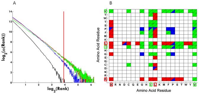
Figure 8. Secondary structural preferences.
(A) The graph shows how the secondary structure assignment changes with rank. Red codes for alpha-helix, green for beta-strand, blue for coil and black for turn. The vertical red line is located at rank 50. (B) Amino acid pairs and their secondary structural location in cells with population ≥50 (rank 50). Color scheme is the same as described for panel (A). Ala, Ile, Leu and Val residues are highlighted with a red or green box.

