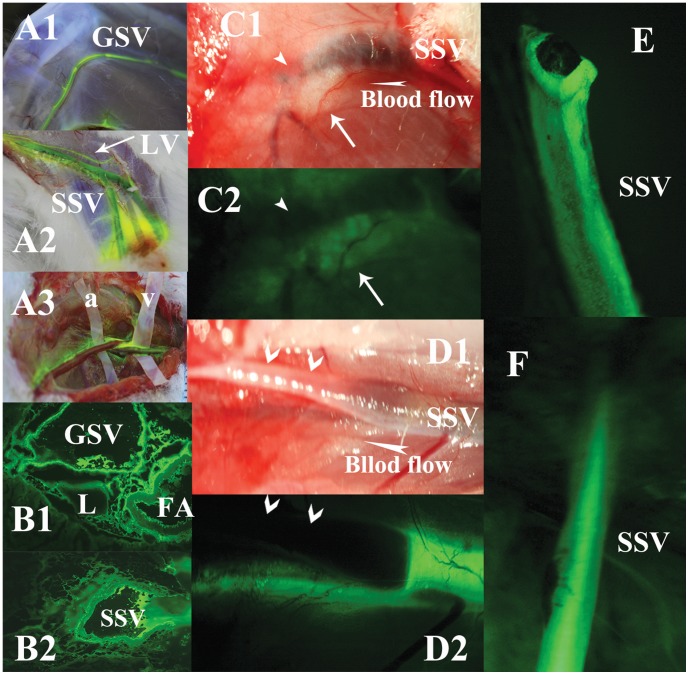
Figure 1. Illustrations of the perivenous tissues, but not artery or intravenous blood, in lower limbs stained by fluorescein from KI3.
The vessels walls of GSV (A1 in the lateral knee), SSV (A2 under the level of popliteal fossa) and a near lymphatic collecting vessel (LV) are stained by fluorescein from KI3, observed in a rabbit of group I. A3 shows venous walls (on plastic spacer v), but not of arteries (on plastic spacer a), are stained in anther rabbit in group I. Observed in group XVI, B1 shows fluorescein accumulates in the interstitial spaces among GSV, FA and LV. B2 shows strong fluorescein around SSV. In group XIII, C1 shows the proximal end of SSV is cut off and ligated (arrowhead points at the broken and ligated end of the SSV, white arrow points at the perivenous loose connective tissues remained due to vasoconstriction after the inside vein is cut off). C2 shows the perivenous tissues are still stained by fluorescein from KI3 even no inside blood vessel. D1 in group VIII shows a segment of SSV, the surrounding tissues on its walls are stripped off and exposed in the air (dried). E2 shows the segmental, unstripped and wet SSV is stained by fluorescein from KI3, Meanwhile, the perivenous loose connective tissues beneath SSV are stained as well. E shows the entire venous walls with broken end are stained by fluorescein from KI3 clearly, isolated from a subject of group II, in contrast to the intravascular blood that is NOT stained. But F shows the intravascular blood in SSV in group XI is stained significantly via intravenous injection.

