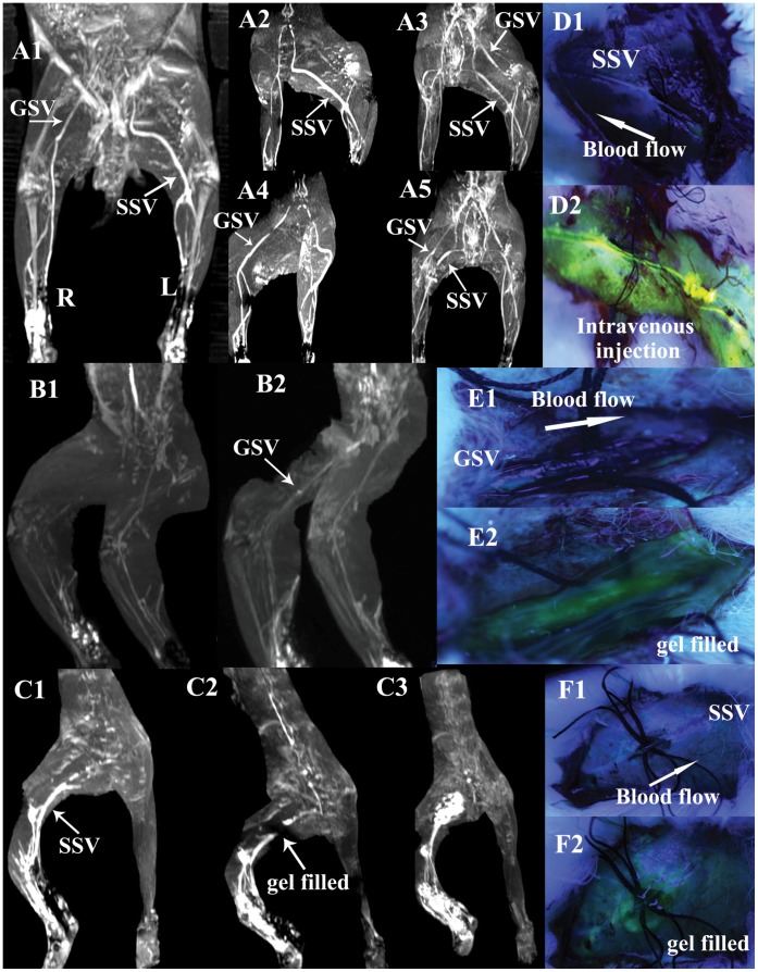
Figure 2. Illustrations of the blockade of the perivenous pathways along GSV or SSV by circular incision and planted sutures, and then restored by transonic gel.
A1–5 show circular incision and planted sutures are performed on segments of left GSV in the medialis knee and right SSV in the lateral thigh in a rabbit of group V. Frontal view (A1) and modified lateral views (A2, A4) show the absence of left “GSV” and right “SSV” scanned at 7 min after KI3 injection in contrast to the intact right “GSV” and left “SSV”. A3, A5, Two views of lower limbs by angiography through intravenous injection via auricular vein, show the interior vascular lumen of either left GSV or right SSV is not blocked by the operations and keep unobstructed actually. D1 shows the perivenous transport of fluorescein sodium from KI3 is blocked by circular incision and planted sutures on SSV in a rabbit of group VI. D2 shows the vascular lumen is actually not obstructed by injecting fluorescein into the vein. B1 shows the blockade in “GSV”, C1 in “SSV” of two rabbits in group VII, E1 in “GSV”, F1 in “SSV” of another two rabbits of group VIII. After filling transonic gel into the incision sites (showed in E2, F2), the centripetal transport of Gd-DTPA or fluorescein sodium from KI3 through perivenous tissues is restored. The entire downstream “GSV or SSV” are visualized again (showed in B2, C2, E2 and F2) (the details of C2 showed in Movie.2). Even the providing-gel is enhanced or stained in a period of time after injection (showed in C3, E2 and F2) (the details of C3 showed in Movie.3).

