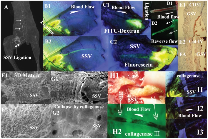
Figure 3. Illustrations of the flow properties of the perivenous pathways exhibited in different groups.
A (a subject in group XII) and B (a subject in group XIII) show the tracers through the perivenous pathways are not due to intravenous blood flow. After the obstruction of a segmental SSV plus its 10 mm surrounding tissues ligated by the sutures, the injected Gd-DTPA in A and fluorescein sodium in B into the proximal side of the ligation can be transported centripetally along the downstream SSV and vertebral vein pointed at by three white arrows. The circle seen in A is water sac due to the injection. B1, B2 are photographed at 5 minutes and 25 minutes respectively after the injection. C1 shows a small amount of FITC-dextran is injected into the perivenous tissues of SSV above the level of popliteal fossa in a subject of group X and photographed at 30 minutes after injection. There are no fluorescent signals found along the vessel walls of the entire length of downstream SSV. C2 shows (photographed at 15 minutes after injection) the perivenous pathway is stained by injected fluorescein sodium into the same position of the same subject. D1 shows the upstream site of a segmental SSV is ligated by suture in group XIII. D2 shows the fluorescein sodium in the downstream site is pushed backwards by repeated massage performance (two white arrows point at the centrifugal pressure direction) and has stained the perivenous pathway reversely. Inmmunostaining results in E1 (×40, CD31), E2 (×20, collagen IV) show no appearances of numerous ring-like groupings or linear structures within the venous adventitia and its surrounding loose connective tissues. F1 (×200),F2 (×500) show porous fiber-bed on adventitia of SSV. G1 (×200), G2 (×10000) show adventitial porous fiber-bed, collapsed, on a segment of SSV hydrolyzed by collagenase type IV. H1 shows a cotton ribbon full of 10% collagenase type III spirals round the venous vessel. H2 demonstrates fluorescein sodium from KI3 is able to bypass the etched gap, be transported along the spiral path and has stained the unetched wall and the downstream venous walls (two arrows point at the positions covered by the cotton ribbon). I1 shows no fluorescein signals on SSV after hydrolyzed by collagenase type I even large amount of fluorescein are injected into the upstream in group XIV. The SSV is drawn aside by spacer to show no fluorescein signals found in surrounding tissues in I2, I3.

