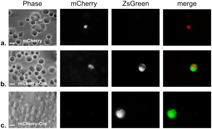
Figure 4. Peritoneal exudate cells from Cre-reporter mice show U-I cells in mice infected with Toxoplasma-mCherry-Cre strain.
Cre-reporter mice that express ZsGreen only after Cre-mediated recombination were inoculated i.p. with 5000 tachyzoites of either the control Pru-mcherry parasites (a) or the Pru-mcherry-Cre parasites (b,c). 4 days after inoculation, the peritoneal exudate cells (PECs) were removed, fixed, and examined by fluorescence microscopy. Panels from left to right show phase, red filter (parasite mCherry), green filter (host ZsGreen), and merge of the red and green filter images, respectively. Note the infected ZsGreen+ cell in (b) and the uninfected ZsGreen+ cell in (c). Scale bars = 10 µm.

