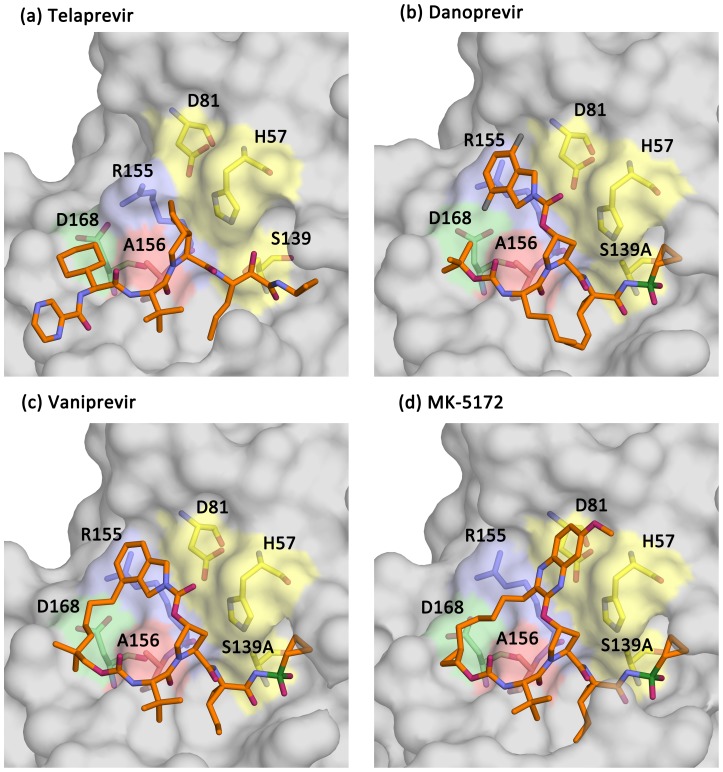
Figure 2. The binding conformations of telaprevir, danoprevir, vaniprevir and MK-5172.
Surface representations of the wild-type protease in complex with (A) telaprevir, (B) danoprevir, (C) vaniprevir, and (D) MK-5172. The catalytic triad is shown in yellow and the R155, A156 and D168 side chains are highlighted in light-blue, pale-green and red, respectively.

