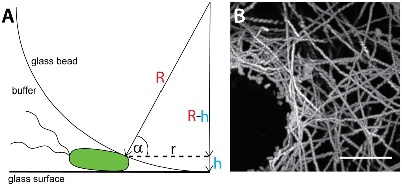
Figure 4. Swimming behavior of S. Typhimurium on gelatine coated glass beads.
Gelatine coated glass beads (500 µm diameter) were placed into a glass-bottom dish and the swimming behavior of mCherry expressing bacteria was recorded by time lapse fluorescence microscopy as described in Fig. 3. (A) Illustration of a bacterium stopping at the glass bottom of a coated glass bead (illustration not to scale). (B) Maximum intensity plot (ImageJ) superimposing all frames of a 15 sec movie acquired at 20 frames per second (300 frames total; suppl. Video S3) illustrating the movement of S. Typhimurium (S.Tmwt(mCherry)) within the vicinity of the bead. Scale bar: 10 µm.

