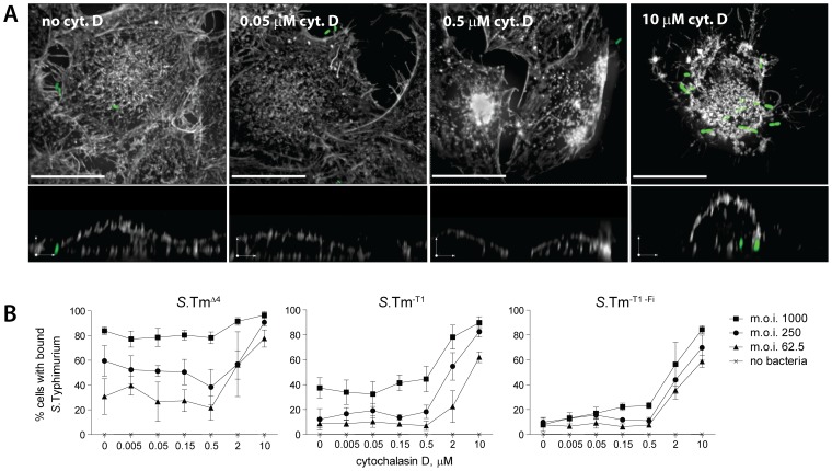
Figure 7. Cellular morphology affects docking of S. Typhimurium.
(A) Cells were incubated with the indicated concentration of cytochalasin D for one hour prior to infection withS.TmΔ4(pGFP) at an m.o.i. of 125 for 10 min. Afterwards, cells were fixed as described in Fig. 6 and stained with DAPI and TRITC-phalloidin (stains actin). Stacks of confocal images were acquired in the actin channel (grey) and the bacteria channel (green). An extended focus projection and a reconstruction of a zx-layer are shown. Scale bar, upper images: 20 µm, zx-layer: 5 µm. (B)Cells were pre-treated with the indicated concentration of cytochalasin D for 1 hour and infected with the respective S. Typhimurium strain for 12 min. After washing, fixing and staining S. Typhimurium docking was quantified by an automated microscopy based docking assay [9]. The curves shown summarize 3 independent experiments. Error bars: standard deviation.

