Fig 3.

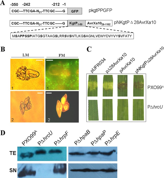
Regulation and secretion of the kgtP gene product. (A) Schematic map showing the kgtP promoter containing the PIP box motifs fused with GFP (top row, in construct pkgtPPGFP), the kgtP promoter, and the N-terminal 50 amino acids of KgtP fused with Δ28AvrXa10 (middle row, in construct pNKgtPΔAvrXa10), and the residue proline (P) and serine (S) consititution of the N-terminal 50 amino acids of KgtP (bottom row). (B) Interaction of X. oryzae pv. oryzae strains with rice suspension cells after a 16-h cocultivation period. Images were acquired by light (LM) or fluorescence (FM) microscopy. X. oryzae pv. oryzae bacterial cells appeared as black dots at rice cell surfaces or gray dots surrounding rice cells under LM and green spindly dots surrounding or near rice cells under FM, while rice cells turned red under FM. The white bar indicates 50 μm. 1, PXO99A(pkgtPPgfp); 2, PΔhrpX(pkgtPPgfp). (C) X. oryzae pv. oryzae strains (3 × 10⁸ CFU/ml) were infiltrated into 2-week-old seedling leaves of IRBB10 rice with needleless syringes. Symptoms were photographed 3 dpi in three independent experiments. The top panels showed symptoms caused by PXO99A(pUFR034), PXO99A(pΔ28AvrX10), PXO99A(pAvrX10), and PXO99A(pNKgtPΔ28AvrXa10), and the bottom panels displayed no symptoms triggered by PΔhrcU(pUFR034), PΔhrcU(pΔ28AvrX10), PΔhrcU(pAvrX10), or PΔhrcU(pNKgtPΔ28AvrXa10). (D) Detection of KgtP secretion by immunoblot analysis. X. oryzae pv. oryzae wild-type PXO99A, PΔhrcU, PΔhrpF, PΔhpaB, PΔhpaP, and PΔhrpE expressing pKgtP-c-Myc were incubated in XOM3 medium. Total protein extracts (TE) and culture supernatants (SN) were analyzed by immunoblotting using anti-c-Myc antibodies.
