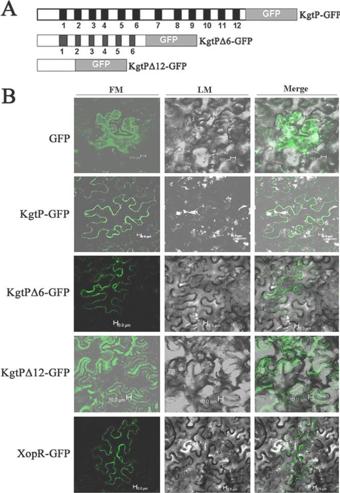
Fig 4.

Subcellular localization of KgtP. (A) Transmembrane distribution and schematic map of deletion constructs of KgtP with GFP. The black boxes indicate the membrane-spanning domains in KgtP protein of X. oryzae pv. oryzae based on the homologous KgtP of E. coli (45, 46). KgtPΔ6 and KgtPΔ12 displayed 6 and 12 membrane-spanning domain deletions, respectively, in the KgtP protein of X. oryzae pv. oryzae. (B) Subcellular localization of KgtP-GFP in tobacco epidermal cells. Expression was driven by the CaMV 35S promoter. For confocal laser scanning microscopy, samples were taken 24 h postinoculation. Images were acquired by light microscopy (LM) or fluorescence microscopy (FM).
