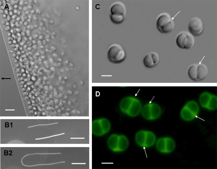
Fig 1.
Morphology and motility of bean-like magnetococci (BMC) based on optical microscopy. Differential interference contrast (DIC) images of BMC are shown in panels A and C. In panel A, the arrow indicates the direction of the magnetic field. For panels B1 and B2, motility tracks of BMC cells in a magnetic field were recorded using dark-field microscopy. BMC cells display a clockwise helical trajectory during forward swimming (B1) and a U-like track in response to reversal of the magnetic field (B2). The exposure time was 3 s for panel B1 and 5 s for panel B2. Panel D shows the fluorescence of BMC exposed to blue light (wavelength 450 to 480 nm). Scale bars, 5 μm (A), 100 μm (B), and 2 μm (C and D).

