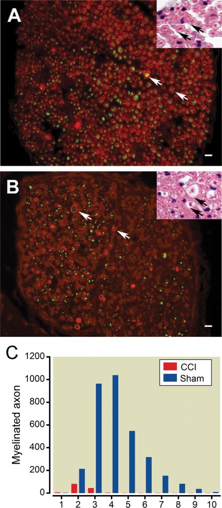
Fig. 3. Morphological alterations produced by chronic constriction injury.
(A) Morphology of the sham control infraorbital nerve (ION): The cytoarchitecture of the ION in the surgical sham control group is shown in nerve cross sections. Axons stained for neurofilament 200 by immunofluorescence (green) are surrounded by myelin sheaths (white arrow) stained for myelin protein zero (red). Inset: Hematoxylin and eosin (H&E) staining shows the normal structure of a fascicle of the infraorbital nerve fiber. Each myelinated nerve fiber axon is densely stained and encircled by the lighter Schwann cell myelin sheath ring (black arrows). (B) Morphology after ION chronic constriction injury (CCI): Most MPZ stained myelin sheaths have lost their circular morphology and axonal staining in the center of their structure, i.e. the immunopositive structures (red) wrapping around the axons (green) have collapsed. Axons are seen scattered in the endoneurium without myelin sheaths or with a few wraps of a damaged myelin sheath (white arrows). Inset: Myelin sheath in infraorbital nerve disrupted by ligation shown 5 weeks after surgery (black arrows). Bars: 10 μm (C) ION size distribution: The histogram shows the infraorbital nerve myelinated axon fiber size distribution and changes after constriction injury.

