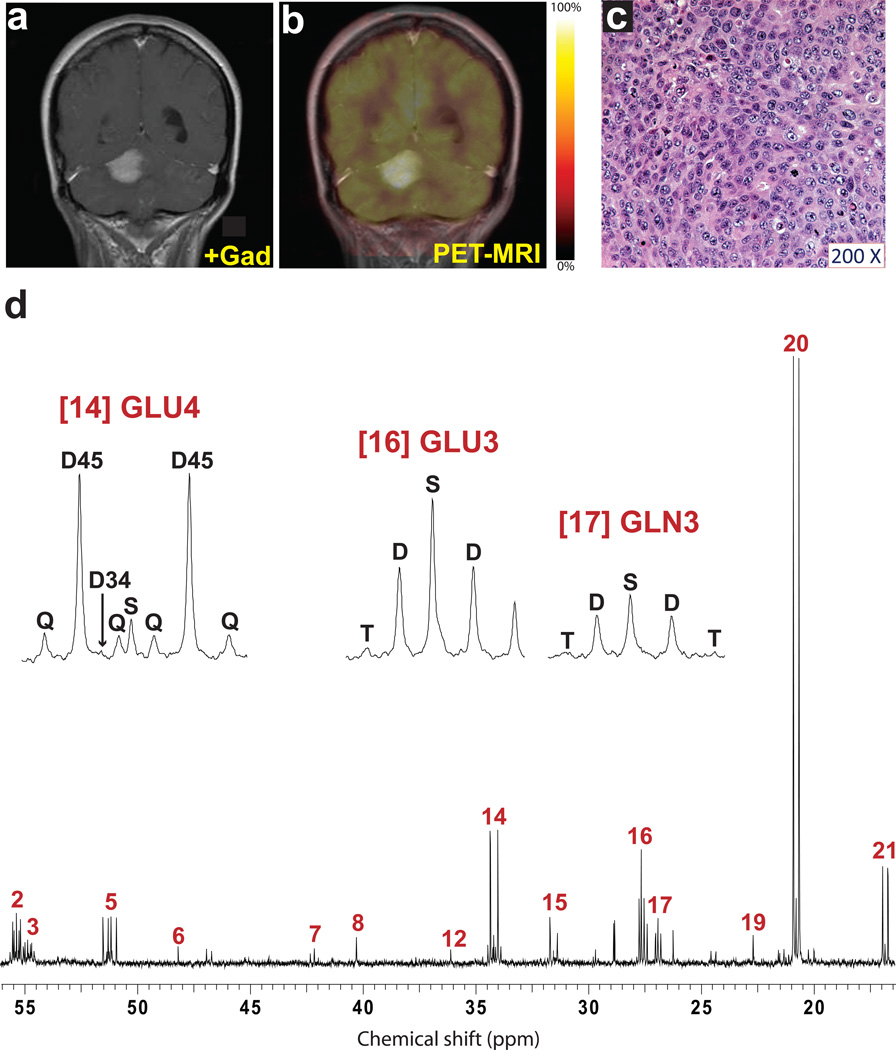
Fig. 6. Tumor glucose metabolism in a patient with metastatic breast cancer.
Pre-operative imaging included (a) T1-weighted post-gadolinium coronal image demonstrating a large, solitary right cerebellar mass and (b) 18FDG-PET scan demonstrating uptake of 18FDG within the mass. (c) Hematoxylin and eosin staining of a histological section prepared from the resected specimen. Immunoperoxidase stains demonstrated expression of mammaglobin and BRST-2 (not shown) in neoplastic cells consistent with origin in the breast. This tumor was positive for the HER-2 oncogene, and was negative for expression of the estrogen and progesterone receptors. Tumor cellularity was >99%. (d) Proton-decoupled 13C NMR spectrum of the tumor. Assignments and abbreviations are the same as in Fig. 1.

