Figure 1.
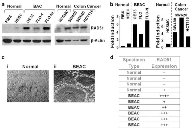
Recombinase (hsRAD51) is overexpressed in Barrett’s esophageal adenocarcinoma (BAC) cell lines and tissue specimens. (a) RAD51 protein expression was analyzed by western blotting in normal human fibroblasts (FIBS), normal primary HEEC, BEAC cell lines (FLO-1, OE33), FLO-1 treated with recombinogen nickel chloride (0.6 mg/ml) for 3 h, normal human colonic smooth muscle cells (HCSMCs) and colonic adenocarcinoma cell lines (SW480, SW620, HCT116). (b) Bar graph showing fold elevation in RAD51 in the adenocarcinoma cell lines of panel (a), relative to control cells. (c) RAD51 immunostaining within BEAC, vs normal tissue. Sections of paraffin-embedded BAC tissues were deparaffinized and treated with anti-RAD51 mouse monoclonal antibody. The samples were then treated with Alexa-Fluor 488-labeled goat anti-mouse secondary antibody and viewed under a fluorescence microscope. The pictures show fluorescence merged with transmitted-light images. (d) Table showing relative expression of RAD51 in all human tissue specimens examined, including those of panel (c). BE, Barrett’s esophagus; BAC Barrett’s adenocarcinoma. A full colour version of this figure is available at the Oncogene journal online.

