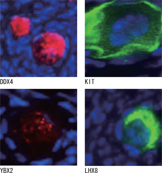
Figure 3. Immunofluorescence analyses for DDX4 (red), KIT (green), YBX2 (red) and LHX8 (green) expression in oocytes within human ovarian cortex tissues.

Sections were counterstained with DAPI (blue) for visualization of nuclei.

Sections were counterstained with DAPI (blue) for visualization of nuclei.