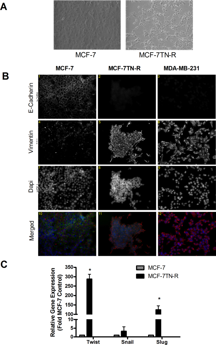
Figure 5. Increased markers of EMT in resistant MCF-7TN-R cells.
(A) MCF-7, MCF-7TN-R, and MDA-MB-231 cells were cultured in eight-well chamber slide for 48 hours. Indirect immunofluorescence was carried out to examine the expression of e-cadherin and vimentin, as described in the Materials and methods section. The nucleus was counter-stained with DAPI. Pseudocolors were assigned as follows: red, E-cadherin; green, vimentin; and blue, nucleus. (B) mRNA gene expression of the EMT markers Twist, Snail and Slug were quantified using qPCR in MCF-7 and MCF-7TN-R cells. Mean values ± SEM of three independent experiments in triplicate are reported.

