
Fig. 3.
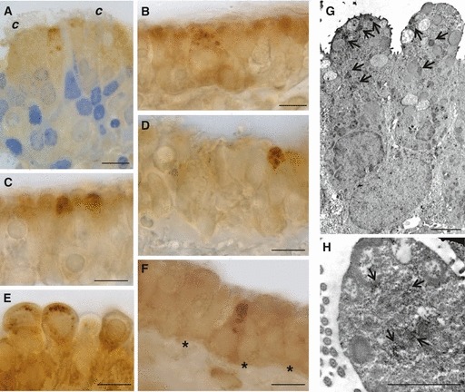
Immunoperoxidase staining showing GLUT5 immunoreactivity at light (A–F) and electron microscopy (G, H) in rat trachea. Immunoreactivity is present in secretory (A–E), and solitary chemosensory cells (F). Nuclei are stained with toluidine blue (A). At ultrastructural level, secretory cells show GLUT5 labeling around or inside the granules (arrow, G), or sparsely distributed in the apical surface (arrow, H). *Basement membrane; c, cilia. Scale bar, 10 μm (A–F), 2000 nm (G, H).