
Figure 4. Photic resetting of the clock is changed in Arvicanthis fed with a HF.
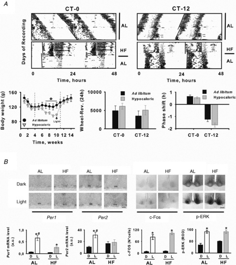
A, top, circadian wheel-running activity from AL and HF Arvicanthis receiving a 30 min light pulse at CT-0 and CT-12. In double-plotted actograms stars indicate the time of light stimulation. Bottom, body weight (left), total locomotion (middle) and group mean (±SEM) phase shifts to light at CT-0 and CT-12 (right) of AL and HF Arvicanthis. In the body weight plot, vertical dotted lines indicate the time in which animals were exposed to HF condition, and arrow indicates body weight values at the moment of light stimulation. B, light exposure induces differential changes on Per1, Per2, c-Fos and p-ERK in the SCN of AL and HF Arvicanthis. Top, representative photomicrographs show coronal sections through the SCN of Arvicanthis kept in constant darkness and exposed to a HF or AL feeding conditions and 30 min light pulse (Light) or a control dark pulse (Dark) at CT-12. Scale bars = 1 mm (Per1–2) and 200 μm (c-Fos, p-ERK). Bottom, semiquantitative analysis of Per1, Per2, c-Fos and p-ERK in the SCN of AL and HF Arvicanthis after 30 min light exposure (L) or after exposure to darkness (D) at CT-12. Data are presented as the mean ± SEM of 4 animals per group. *P < 0.05, differences between light conditions (Light vs. Dark). #P < 0.05, differences between groups (AL vs. HF).