


Figure 2.
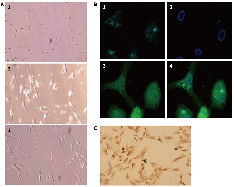
Isolation, culture, and identification of primary rat hepatic stellate cells. A: Primary rat hepatic stellate cells (HSCs) on days 1, 2, and 5 after isolation and plated in culture; B: HSCs isolated from Sprague-Dawley rats showed characteristic multiple lipid droplets with a rapid fade of the blue-green autofluorescence at 328 nm. 1: Lipid droplets; 2: Cell nucleus; 3: Cell body. 4: Merged images; C: HSC culture after the first passage and stained with anti-α-smooth muscle actin antibody.