FIG. 1. .
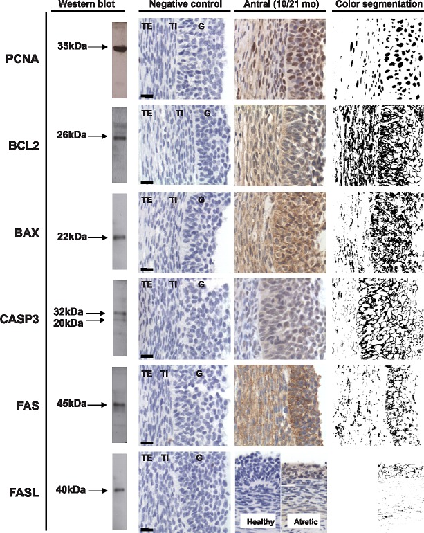
Representative images of PCNA, BCL2, BAX, CASP3, FAS, and FASL immunostaining in antral follicles are shown in the third column. Verification of antibody specificity by Western blot analyses of ovarian homogenate and negative controls for immunostaining demonstrating the specificity of the antibody are shown in the left two columns, respectively. The fourth column shows segmentation analyses results of immunostaining. G, granulosa cells; TE, theca externa; TI, theca interna. Bar = 25 μm.

