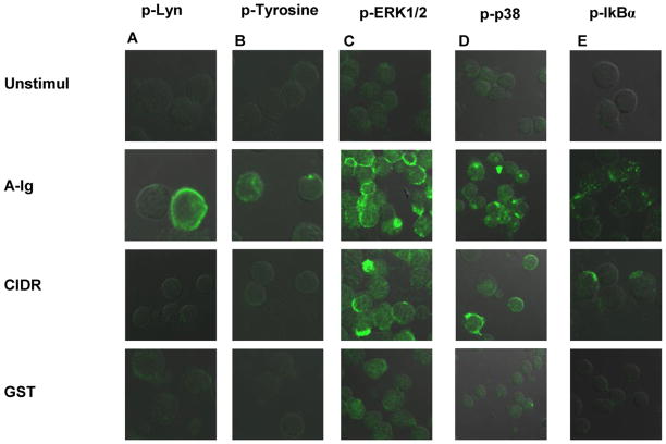
Fig. 4.
PfEMP1–CIDR1α does not induce Lyn and Tyrosine phosphorylation but triggers the phosphorylation of ERK1/2, p38 and IkBα. (A) CD19+ B cells were either untreated or stimulated for 2 min with either anti-Ig F(ab′)2 10 μg/mL or PfEMP1–CIDR1α 100 μg/mL and GST 50 μg/mL as control. In each case the cells were fixed, permeabilized, and stained with primary rabbit antibody specific for phospho-Lyn while the secondary goat anti-rabbit antibody was labelled with Alexa-488. (B–D) The same as in (A), except for the stimulation times: 3, 10 and 60 min, respectively, and the specific primary rabbit antibody for phospho-tyrosine used. (E) CD19+ B cells were stimulated as above for 30 min and stained with specific primary mouse antibody for phospho-IkBα. The secondary goat anti-mouse antibody was labelled with Alexa-488. Representative confocal images of Alexa-488 are shown; approximately 200 cells were analyzed from three independent experiments for each kinase. A and B present a doubled magnification compared with C, D and E.

