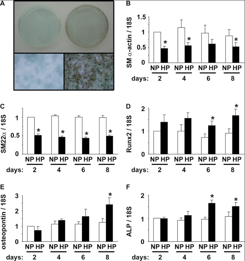
FIGURE 1.
High phosphate concentration induced calcification of SMCs. A, rat aortic SMCs were cultured with normal (left) or high (right) phosphate concentration for 10 days. Representative pictures of von Kossa staining are shown (n = 3). Top, appearance of 60-mm dishes. Bottom, magnification x100. B–F, SMCs were incubated with normal (NP) or high (HP) phosphate concentration for 2, 4, 6, and 8 days. Expression of SM α-actin (B), SM22α (C), Runx2 (D), osteopontin (E), and ALP (F) mRNA was determined by real-time RT-PCR. Values represent the means ± S.E. *, p < 0.05 compared with SMCs with normal phosphate medium (n = 4).

