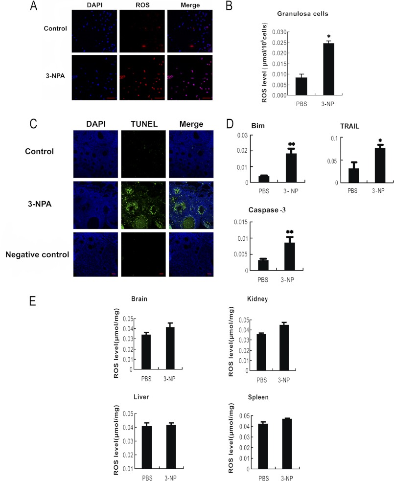
FIGURE 1.
Oxidative stress-induced apoptosis in MGCs. Mice were intraperitoneally injected with PBS or oxidant 3-NP, respectively. The intracellular ROS in the harvested follicular MGCs (A) and from the TUNEL assay of follicular MGCs in the ovary sections (C) from intraperitoneally injected mice were measured with a microscope. Bar, 50 μm. B and E, ROS levels in follicular MGCs and several tissues were also quantified by nitro blue tetrazolium (NBT) staining. D, qRT-PCR showed the induction of apoptosis-related genes in response to oxidant treatments; the relative expression data were normalized to the amount of cellular β-actin. The corresponding data above represent mean ± S.E. (error bars) (n = 3). The statistical significance between groups was analyzed by one-way ANOVA. *, p < 0.05; **, p < 0.01.

