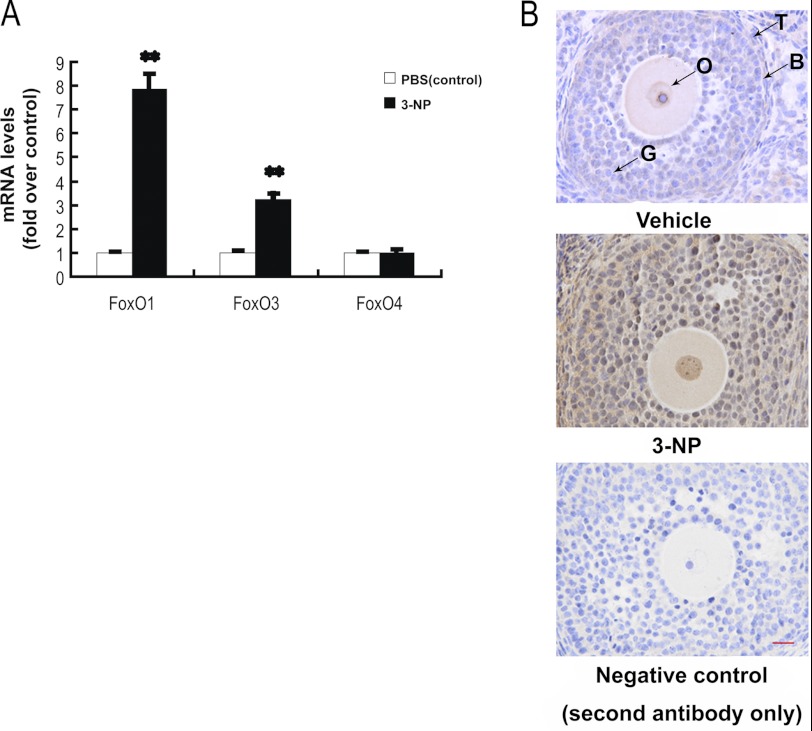
FIGURE 2.
Oxidative stress up-regulates FoxO1 expression in follicular MGCs. A, qRT-PCR showed mRNA transcription changes of FoxO members in response to oxidative stress in follicular MGCs. Data are mean ± S.E. (error bars) (n = 3). The relative expression data were normalized to the amount of cellular β-actin. The statistical significance between groups was analyzed by one-way ANOVA. *, p < 0.05; **, p < 0.01. B, immunostaining of granulosa cells in ovary sections was detected by using anti-FoxO1 as described under “Experimental Procedures.” Bar, 20 μm. O, oocyte; G, granulosa cells; B, basement membrane; T, theca cells.

