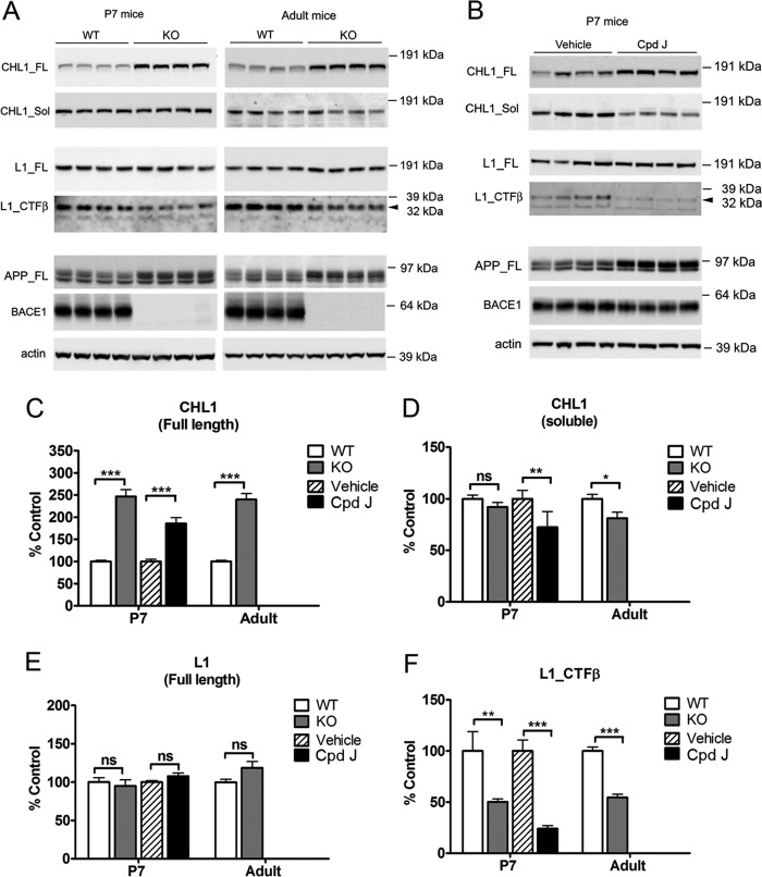
FIGURE 3.
Validation of CHL1 and L1 as BACE1 substrates in mouse brain homogenates from both genetic and acute pharmacological inhibition systems. A, representative Western blots of brain homogenates from 7-day-old (P7) or adult BACE1 knock-out mice and wild type control mice or (B) P7 wild type mice acutely treated with BACE1 inhibitor compound J (Cpd J) or vehicle (as control). Brain hemispheres were sequentially homogenized in TBS buffer and in RIPA buffer to prepare TBS-soluble and RIPA lysate fractions. The soluble fragment of CHL1 (CHL1_Sol) was detected from TBS-soluble fraction, and the full-length proteins (CHL1_FL and L1_FL) and the β-cleaved C-terminal fragment of L1 (L1_CTFβ) were detected from the RIPA fraction. C–F, semi-quantification of the Western blots. Data were mean ± S.E., n = 8 mice for each group, Student's t test (*, p < 0.05; **, p < 0.01; ***, p < 0.0001; ns, statistically not significant).

