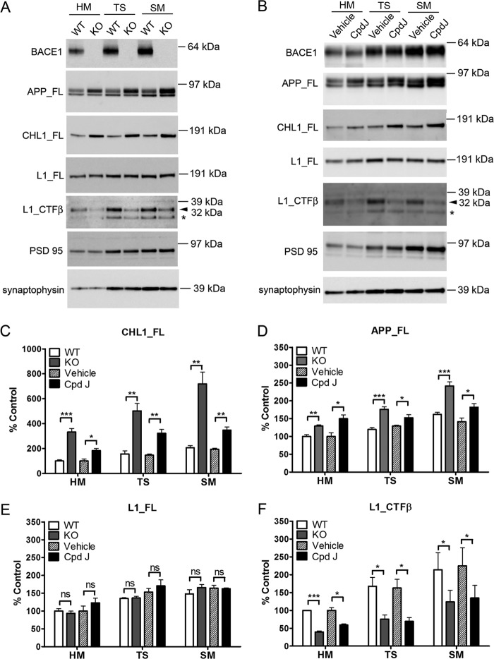
FIGURE 4.
Validation of CHL1 and L1 as BACE1 substrates in mouse brain synaptic fractions using both genetic deletion and acute pharmacological inhibition. A, representative Western blots of brain homogenate (HM), total synaptosome (TS), and synaptic membrane (SM) fractions from 7-day-old BACE1 knock-out mice or wild type control mice, or B, P7 WT mice acutely treated with BACE1 inhibitor compound J or vehicle. PSD-95 and synaptophysin were detected as synaptic markers. Notice that the L1 CTFβ (32 kDa) and a shorter L1 CTF fragment (*, released by other protease than BACE1) were indicated. C–F, semi-quantification of the Western blots. Data were mean ± S.E., n = 4 for each group, Student's t test (*, p < 0.05; **, p < 0.01; ***, p < 0.0001; ns, statistically not significant).

