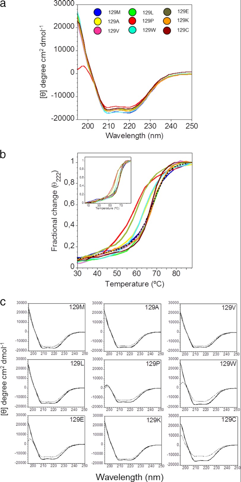
FIGURE 1.
Secondary structure analysis by circular dichroism. a, far UV-circular dichroism spectra of native HuPrP(90–231), mutants in position 129 at 4 °C. The color coding for mutant identification is shown in the inset. b, thermal unfolding curves of 129-mutants monitored at 222 nm. The colors are as shown in Fig. 1a. The dashed spectrum shows the thermal unfolding curve of WT HuPrP(121–231). c, far UV-circular dichroism spectra of mutants in position 129 at 4 °C before (solid line) and after (dashed line) one thermal unfolding/refolding.

