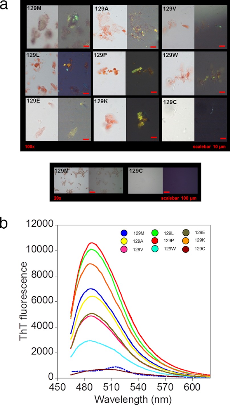
FIGURE 2.
Amyloid fibril formation of HuPrP(90–231) mutants. a, micrographs of Congo red-stained aggregates of HuPrP(90–231), mutants in position 129 formed under native conditions in 2-ml cryotubes. Shown are open (left image) and crossed polarizers (right image). Scale bar, 10 μm. All mutants except for M129C displayed Congo red birefringence. A side-by-side overview comparison at lower magnification of WT (129M) and M129C is shown in the lower panel to highlight the lack of Congo red-stained aggregates from M129C. b, thioflavin T fluorescence spectra of aggregates of HuPrP(90–231), mutants in position 129 formed under native conditions. The color coding is shown in the inset. The dashed spectrum shows the fluorescence for native HuPrP(90–231) in the presence of ThT.

