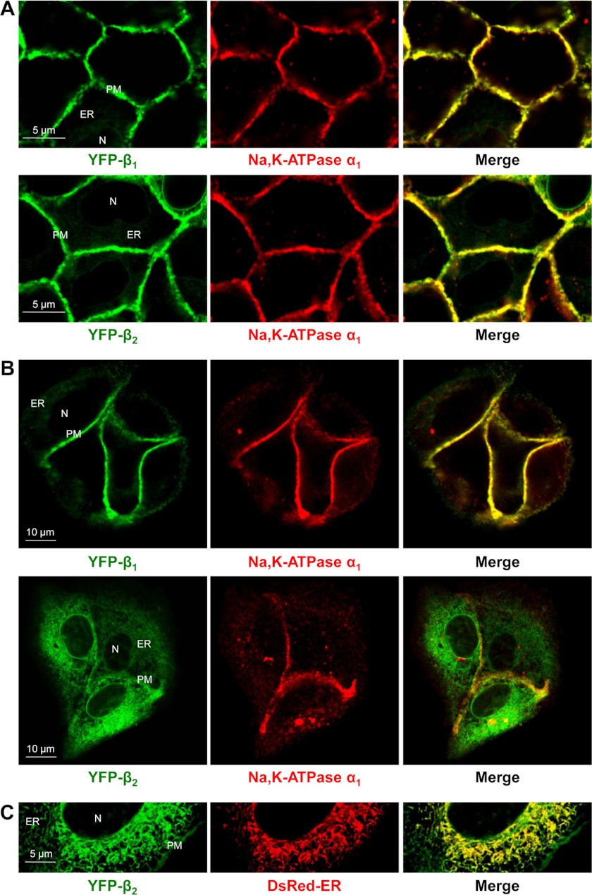
FIGURE 5.
The amount of α1-unbound YFP-β2 retained in the ER of MDCK cells is greater than that of YFP-β1. Horizontal confocal microscopy sections of confluent monolayers (A) or dispersed colonies (B and C) of MDCK cells expressing either YFP-β1 or YFP-β2. Both YFP-β1 and YFP-β2 (green) are co-localized with the endogenous α1 subunit (red) in the lateral membranes, but not inside the cells as detected by immunostaining of fixed cells using the monoclonal antibody against the Na,K-ATPase α1 subunit (A and B). The intracellular retention of α1-unassembled YFP-β2 is more prominent than that of YFP-β1 and more evident in dispersed colonies than in confluent monolayers. This intracellular fraction of YFP-β2 (green) shows co-localization with the ER (red) as detected by transient expression of the fluorescent ER marker, DsRed2-ER (C). N, nucleous; PM, plasma membrane.

