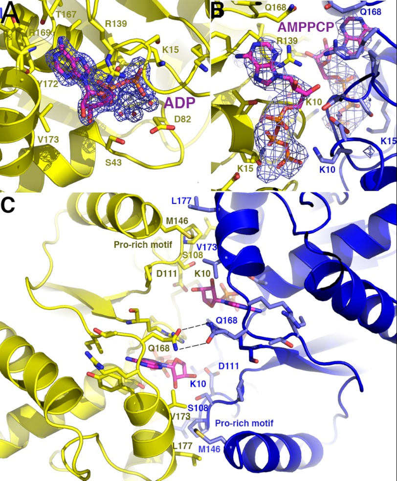
FIGURE 3.
Close-up of the ParF adenine nucleotide-binding pocket. A, view of the adenine nucleotide pocket in the ParF-ADP structure. Residues that make key interactions with the ADP are shown as sticks. Also shown is an Fo − Fc omit map (contoured at 5σ) in which the ADP and hydrated magnesium ion were omitted. B, close-up of the nucleotide-binding pocket of the dimeric ParF-AMPPCP structure. Residues that contact the AMPPCP are shown as sticks and colored according to the subunit. Superimposed is the omit electron density map (blue mesh and contoured at 4σ) in which the AMPPCP molecules and magnesium ions were omitted. C, close-up showing cross-contacts between the two ParF subunits that stabilize the nucleotide sandwich dimer.

