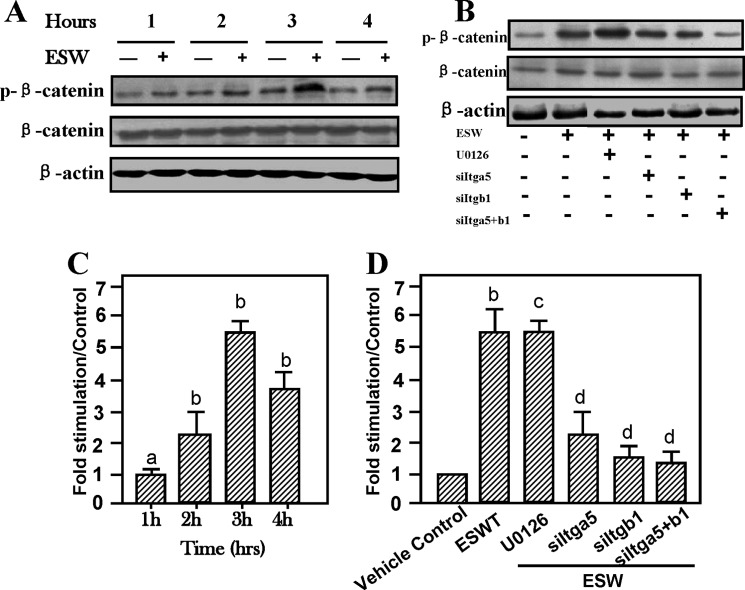
FIGURE 6.
Enhancement of β-catenin activity by ESW (500 impulses at 10 kV) stimulation after elevation of integrin α5 and β1 expression. ESW raised β-catenin phosphorylation in 3 h (A). ERK1/2 inhibitor U0126 did not alter the activation of β-catenin (B). Cytosolic extracts of osteoblasts treated with ESW in the presence of U0126 (with a final concentration of 20 μm) for 60 min prior to ESW were subjected to Western blotting. Phosphorylated β-catenin and β-catenin were probed with anti-phospho-β-catenin and β-catenin primary monoclonal antibodies, respectively. C, note that in comparison with the control, ESW exposure markedly elevated the activation of β-catenin. Further studies on the relationship between expression of integrins and β-catenin by transfection have shown that knocking out integrins led to base-line level expression of activation of β-catenin (B and D). D, summary of the results (mean ± S.E. (error bars), n = 4, triplicate in each experiment). a, p > 0.05 as compared with the control at the same period. b, p < 0.01 as compared with the control at the same period. c, p > 0.05 as compared with the ESW group. d, p < 0.01 as compared with the ESW group.

