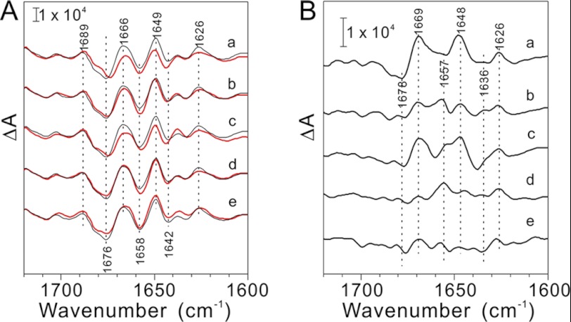
FIGURE 4.
A, S2QA−/S1QA FTIR difference spectra in the amide I region (1700–1600 cm−1) of the NaCl-washed (a, red line) and PsbP-reconstituted (b–e, red lines) PSII membranes. The spectra of the untreated PSII particles are shown in black. PSII particles were reconstituted with WT (b), H144A (c), D165V (d), and H144A/D165V (e) PsbP proteins, respectively. B, untreated minus NaCl-washed (a) or PsbP-reconstituted (b–e) double difference spectra of S2QA−/S1QA FTIR spectra in the amide I region. b–e: WT, H144A, D165V, and H144A/D165V-reconstituted PSII, respectively.

