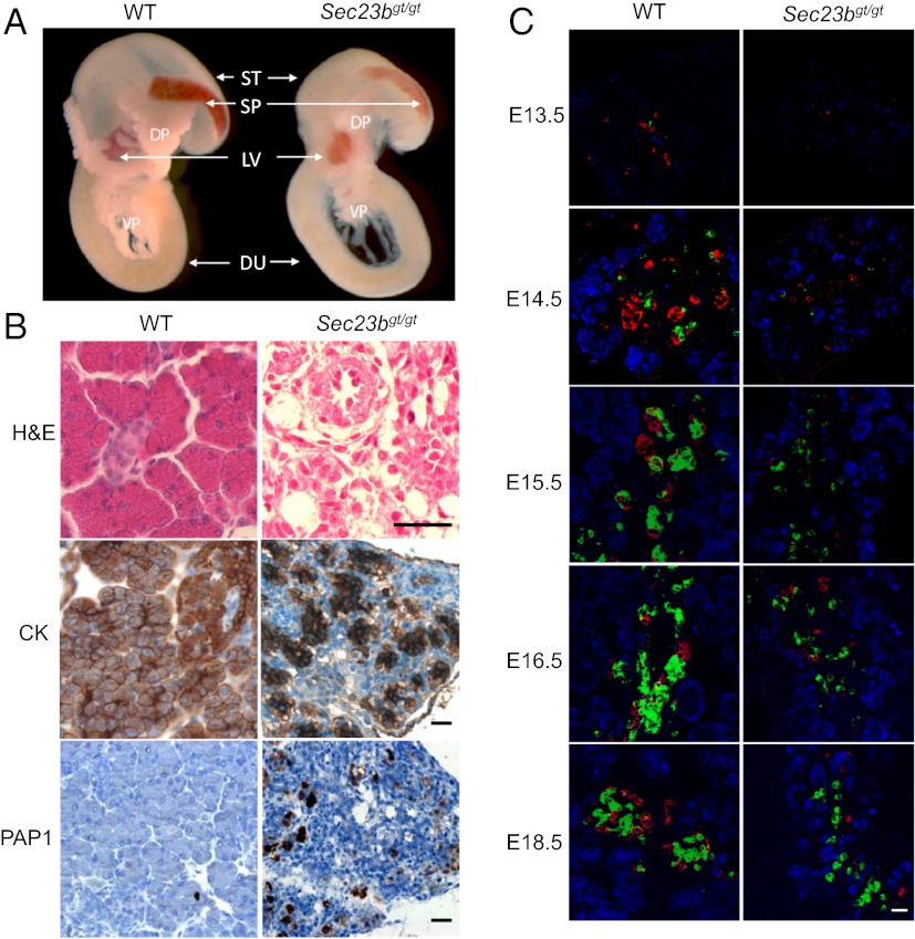
Fig. 2.
Pancreatic defect in Sec23bgt/gt mice. (A) Pancreas and adjacent tissues dissected from E18.5 embryos. Pancreatic tissues from Sec23bgt/gt embryos are smaller and less demarcated than in WT embryos. DP dorsal pancreas; DU, duodenum; LV, liver; SP, spleen; ST, stomach; VP, ventral pancreas. (B) H&E and immunohistochemical staining of pancreas from WT and Sec23bgt/gt mice. PAP1, pancreatitis associated protein 1; CK, cytokeratin. (Scale bar: 30 μm.) (C) Immunofluorescence staining of pancreas at E13.5, E14.5, E15.5, E16.5, and E18.5. Cryosections were costained with rabbit anti-carboxylpeptidase A (blue) for exocrine cells and mouse anti-glucagon (red) and guinea pig anti-insulin (green) for α- and β-endocrine cells, respectively. (Scale bar: 25 μm.)

