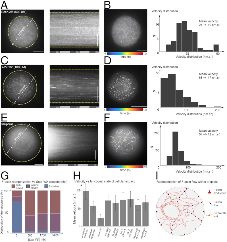
Fig. 4.
Testing the role of the spatial localization of F-actin nucleation sites, the requirement of myosin II, and the regulation of the cell cycle in the generation of symmetry breaking and actin flow. (A) Kymograph of F-actin dynamics within droplet in presence of Scar-WA (500 nM). (B) Velocity field in presence of Scar-WA inhibitor (500 nM) and velocity distribution (N = 3 droplets, approximately 70 trajectories, mean = 21 ± 10 nm). (C) Kymograph of F-actin network in presence of Rho-Kinase inhibitor Y27632 (100 μM). (D) Velocity field of F-actin network dynamics in presence of Rho-Kinase inhibitor Y27632 (100 μM) and velocity distribution (N = 5 droplets, approximately 100 trajectories, mean = 68 ± 17 nm). (E) Kymograph of F-actin dynamics within droplet in interphase state. (F) Velocity field in interphase state and velocity distribution (N = 10 droplets, approximately 500 trajectories, mean = 54 ± 12 nm). (G) Histogram of events characterizing the network dynamic state for increasing concentration of Scar-WA (0, 0.5, 1.7, and 5 μM). (H) Histogram of the mean velocity as a function of the functional state of confined cellular extract. (I) Schematic of F-actin-based structure and flow within droplet. Scale bars, 10 μm.

