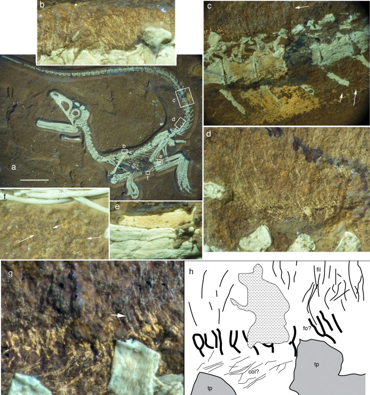
Fig. 3.
Soft tissue preservation in Sciurumimus. (A) Overview of skeleton under UV light, with position of magnifications in B–F indicated. (Scale bar: 50 mm.) (B) Fine filaments above the scapular region of the dorsal vertebral column. (C) Anterior midcaudal section with long dorsal filaments (upper white arrow), preserved skin (yellow patch), and fine filaments at the ventral lateral tail flank (lower white arrows). (D) Long filaments, anchored in the skin at the dorsal tail base. (E) Small section of possibly fossilized muscle tissue along the posterior edge of the tibia. (F) Small, fine filaments ventral to the gastralia in the abdominal area (arrows point to individual filaments). (G) Magnification of soft tissues dorsal to the ninth and 10th caudal vertebra. (H) Interpretative drawing showing possible follicles. Greenish white structures are bone, fine greenish lines above the vertebrae are preserved filaments, and yellow parts represent skin structures. Arrow in G points to a filament entering one of the vertical skin structures that might represent follicles. col, collagen fibers in the skin; fil, filaments; fo? possible follicles; tp, transverse process. All photographs were taken under UV light.

