Fig. 3.
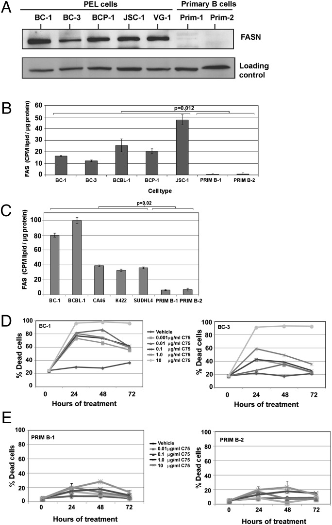
FAS is a critical and essential metabolic pathway for the proliferation of PEL. (A) PEL express higher levels of FASN compared with primary B cells. Ku80 is shown as a loading control. (B) The rate of FAS in PEL is significantly higher (P = 0.012) compared with primary B cells. (C) PEL (BC-1 and BCBL-1) have higher FAS rates compared with BL (CA46) and FL (K422 and SUDHL4). Collectively, all B-NHL have a higher FAS rate compared with primary B cells. Data are normalized to total input protein and is one representative of multiple independent experiments. Error bars are ± SEM. (D) Inhibition of FAS using varying concentrations of the FASN inhibitor, C75, leads to a dose-dependent increase in cell death in BC-1 (Left) and BC-3 (Right) PEL cells, as measured by trypan blue exclusion. (E) Inhibition of FAS using C75 leads to minimal cell death in primary B cells from two donors, as measured by trypan blue exclusion. Error bars are ± SEM.

